
Core facilities Acıbadem Mehmet Ali Aydınlar University
Electron Microscopy Laboratory
FEG-SEM and 120 kV TEM imaging down to 1 nm, with EDS elemental analysis and correlative MAPS imaging.
What we offer
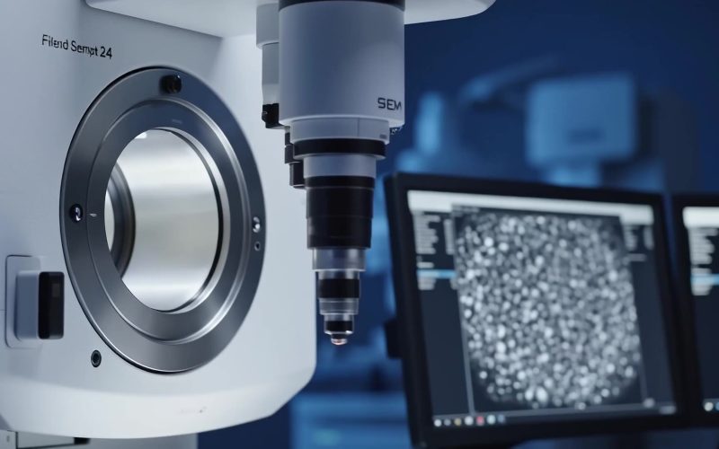
Imaging of micro- and nanostructures is an indispensable part of R&D. With the field-emission scanning electron microscope (FEG-SEM) in our Electron Microscopy Laboratory, materials can be imaged at the micro and nano scale and their elemental composition determined by energy-dispersive X-ray spectroscopy (EDS). Depending on the sample, images can be taken at resolutions down to 1.0 nm and magnifications up to 1,000,000x.
The system's navigation camera captures a colour image of the whole stage and stubs with the samples, so the region of interest is found quickly. The MAPS correlation feature links images from other microscopes (fluorescence, light, confocal, IR and Raman), so regions marked with those techniques can be imaged at high magnification with the SE and BSE detectors. Three analysis modes cover conductive, insulating and biological or moist samples.
The 120 kV transmission electron microscope takes ultra-thin tissue sections prepared with the ultramicrotome, nanoparticles, graphene and similar samples, and its C-TWIN lens technology gives the highest contrast. The laboratory is used for the morphological assessment of tissues and organs, hair samples, tissue engineering, cell and tissue culture, bacteria and viruses, and in the diagnosis of some diseases.
-
SEM imaging
High-vacuum, low-vacuum and environmental (ESEM) modes for conductive, uncoated insulating and biological samples; 1.0–1.3 nm at 30 kV.
-
EDS elemental analysis
Energy-dispersive X-ray spectroscopy of the imaged region.
-
TEM imaging
120 kV transmission imaging of ultra-thin sections, nanoparticles and graphene, 25–650 kx.
-
Correlative imaging
MAPS correlation of regions marked on fluorescence, light, confocal or Raman microscopes.
Research themes served
Equipment 2
- Scanning electron microscope Thermo Fisher Scientific (FEI) Quattro S ESEM, FEG Imaging to 1.0 nm at 30 kV and 1,000,000x; high-vacuum, low-vacuum and ESEM modes; EDS; navigation camera; MAPS correlation.
- Transmission electron microscope Thermo Fisher Scientific (FEI) Talos L120C, 120 kV Line resolution 0.204 nm, point resolution under 0.37 nm, 25–650 kx, alpha tilt −90° to +90°; C-TWIN lens.
Access terms & pricing
The laboratory is part of the Test and Analysis Unit (ACUTAB) and is open to university and industry researchers. Requests are made with the ACUTAB test list and application forms; analyses are carried out and reported to national and international standards.
Prices on request through ACUTAB.
Facility enquiry
Enquire about EM laboratory
Tell us what you would like to do. Your message goes to the Global Research, Reputation and Rankings Office.
- Send your enquiryFill in the short form.
- GR3 routes itLogged and passed to the right group or facility.
- We reply by e-mailWith next steps, terms or a contact person.